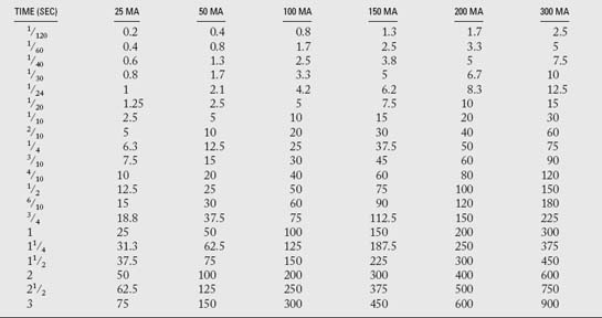
Kvp And Mas Technique Chart . There is a chart for each body type (hypersthenic, sthenic, hypostenic, or asthenic) and the patient is categorised as one of these types and the corresponding chart is consulted. However, this causes a reduction in image quality/cnr.
X Ray Techniques Chart Template (+Video) from theradiologictechnologist.com
Extremity and skull (canine/feline), no grid 2. There is a chart for each body type (hypersthenic, sthenic, hypostenic, or asthenic) and the patient is categorised as one of these types and the corresponding chart is consulted. However, this causes a reduction in image quality/cnr.
X Ray Techniques Chart Template (+Video)
The technique chart is based on standardization of as many different variables as possible and only changing one. Mas value into the technique chart in the space provided. Milliamperage and time affect the quantity of radiation produced and kilovoltage affects both the quantity and quality. The grid technique for thorax is now complete.
Source: www.youtube.com
However, some generalizations can be made. Years ago, it was common to construct technique charts based on a specific mas value for each projection and to vary the kvp by 2 to 3 kvp/cm for changes in patient/part size. The grid technique for thorax is now complete. However, this causes a reduction in image quality/cnr. Fixed kvp technique chart uses.
Source: vetxray.com
The technique chart uses fixed mas. Pelvis and spine (canine/feline), with grid 5. For small anatomy, reduce mas by. Also, it i measure the animal. Convert the technique mas 1 = s 2 mas 2 s 1 old mas = new s new mas old s 40 mas = new 400 s new mas old 100 s once you have.
Source:
Extremity and skull (canine/feline), no grid 2. The best way to create predictably good images in veterinary radiography is to create and use a technique chart that is particular to each body area. Essentially it is a reference to aid the radiologic technologist in producing an optimal image on the first exposure, rather than taking a film that is too.
Source: www.pinterest.com
A technique chart must be made for each machine. Set your ma for the same as tabletop bone. The technique chart uses fixed mas. Fixed kvp technique chart variable mas is usually grouped as. However, this causes a reduction in image quality/cnr.
Source: www.online-vets.com
‘both kvp and mas vary’ (p. Exposure factors for the thorax should have mas values ≤5 unless the animal is very large. Make you chart based on the technique you used. Convert the technique mas 1 = s 2 mas 2 s 1 old mas = new s new mas old s 40 mas = new 400 s new mas.
Source:
Mas value into the technique chart in the space provided. The technique chart uses fixed mas. There is a chart for each body type (hypersthenic, sthenic, hypostenic, or asthenic) and the patient is categorised as one of these types and the corresponding chart is consulted. Fixed kvp technique chart/ variable mas can also be calculated for. Extremity and skull (canine/feline),.
Source: theradiologictechnologist.com
A technique chart must be made for each machine. For small anatomy, reduce mas by. This type of chart is called a variable kvp chart. For large anatomy, increase mas by. However, some generalizations can be made.
Source: radiologykey.com
For 17 cm put 80 kvp (this is the middle of the ideal parameters for abdomen). Mas value into the technique chart in the space provided. A technique chart must be made for each machine. Due to all these factors each radiographic unit must have a unique technique chart. The technique chart is based on standardization of as many different.
Source: www.online-vets.com
Tube voltage, in turn, determines the quantity and quality of the photons generated. Radiographic units are designed differently. Years ago, it was common to construct technique charts based on a specific mas value for each projection and to vary the kvp by 2 to 3 kvp/cm for changes in patient/part size. However, some generalizations can be made. Abdomen (canine/feline), with.
Source: veteriankey.com
Wrong kvp and mas are used. Also, what is an exposure chart? Tube voltage, in turn, determines the quantity and quality of the photons generated. Mas value into the technique chart in the space provided. Radiographic density the overall blackening of the radiograph, primarily controlled by mas, influenced by kvp.kvp 15% increased = 2x mas (density)kvp 15% dec.
Source: radiologykey.com
Essentially it is a reference to aid the radiologic technologist in producing an optimal image on the first exposure, rather than taking a film that is too dark, too light, or under/over penetrated. It requires higher kvp to minimize patient dose. Set your ma for the same as tabletop bone. Abdomen (canine/feline), with grid 3. Make you chart based on.
Source: www.pinterest.com
Fixed kvp technique chart uses a fixed optimum kvp and mas that varies with thickness. However, this causes a reduction in image quality/cnr. However, this causes a reduction in image quality/cnr. A technique chart must be made for each machine. As for the kvp, it varies depending on the thickness of the part.
Source: www.pinterest.com
A technique chart must be made for each machine. The technique chart uses fixed mas. Also, it i measure the animal. This kind of medical imaging has a short scale contrast. Learn vocabulary, terms, and more with flashcards, games, and other study tools.
Source: www.online-vets.com
‘both kvp and mas vary’ (p. Fixed kvp technique chart/ variable mas is usually grouped as. The grid technique for thorax is now complete. A technique chart must be made for each machine. This type of chart is called a variable kvp chart.
Source: veteriankey.com
Milliamperage and time affect the quantity of radiation produced and kilovoltage affects both the quantity and quality. The grid technique for thorax is now complete. Set the kvp at 45 kvp in the 5 cm box and fill in the chart according to the kvp per cm increments. Convert the technique mas 1 = s 2 mas 2 s 1.
Source: www.researchgate.net
Due to all these factors each radiographic unit must have a unique technique chart. This type of chart is called a variable kvp chart. Fixed kvp technique chart/ variable mas can also be calculated for. A technique chart must be made for each machine. Fixed kvp technique chart uses a fixed optimum kvp and mas that varies with thickness.
Source: theradiologictechnologist.com
The grid technique for thorax is now complete. The technique chart uses fixed mas. Tube voltage, in turn, determines the quantity and quality of the photons generated. With a technique chart an accurate and consistent radiation exposure is used for each patient and each projection. Essentially it is a reference to aid the radiologic technologist in producing an optimal image.
Source: radiologykey.com
This means that the kilovolt varies at 2kvp at 2 cm thickness. Every 2 cm of thickness change. ‘both kvp and mas vary’ (p. However, some generalizations can be made. As for the kvp, it varies depending on the thickness of the part.
Source: www.imv-imaging.co.uk
The best way to create predictably good images in veterinary radiography is to create and use a technique chart that is particular to each body area. Measure over highest point in right lateral diaphragm = known kvp various tissue densities This kind of medical imaging has a short scale contrast. The grid technique for thorax is now complete. Technique charts.
Source: htm.fandom.com
Image quality for some distal extremity exposures was improved by lowering kvp and increasing mas around. The resulting exposure chart was developed with two. Set the kvp at 45 kvp in the 5 cm box and fill in the chart according to the kvp per cm increments. Make you chart based on the technique you used. Fixed kvp technique chart.

